
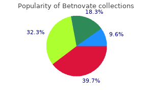
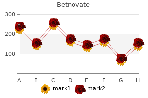
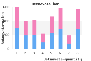
Betnovate

ChristopherWalters, MD
- Cardiology Fellow
- Gill Heart Institute
- Division of Cardiovascular Medicine
- University of Kentucky
- Lexington, Kentucky
Left: Major macular involvement suggests poor visual prognosis after cataract removal acne red marks order betnovate 20gm with mastercard. Right: Minor macular involvement indicates better visual acuity after cataract removal acne 8 weeks pregnant buy line betnovate. Visual acuity outcomes with and without surgery in patients with persistent fetal vasculature acne 5 weeks pregnant cost of betnovate. Oph unilateral congenital cataract caused by persistent fetal vasculature or minimal fetal thalmology acne face mask purchase betnovate from india. Premature infants (poor absorption and ^ requirement of zinc) when weaned off breast milk (which has adequate zinc level) 2. The Hospital Juvenile Hyaline Fibromatosis for Sick Children: Atlas of Pediatrics. Vascular Tumors Vascular Malformations Infantile and Congenital Capillary Malformation (slow ow): Hemangiomas Port-Wine Stain (Nevus Flammeus) Kaposiform Venous Malformation (slow ow): Hemangioendothelioma Cavernous Hemangioma, Phlebectasia Pyogenic Granuloma Lymphatic Malformation (slow ow): Lymphangioma (Lymphangioma Circumscriptum Cystic Hygroma, Cavernous Lymphangioma) Tufted Angioma Arteriovenous Malformation (fast ow): Cirsoid Aneursm Congenital Combined Malformation (slow or fast ow) Hemangiopericytoma A. Paul Getz) B: Anhidrotic ectodermal dysplasia D: Palmoplantar keratoderma in Vohwinkel syndrome (Courtesy of Dr. Subcutaneous fat necrosis of the newborn: a case report and review of the literature. She underwent eight treatment sessions with the Hospital Materno Infantil a mean interval of 30 days between each session. The study was conducted at the Celia Kalil Presidente Vargas and responsible Dermatology Clinic. Results: the port wine stains disappeared completely with a very low for the Cosmiatry Outpatient Clinic of the Santa Casa de Misericordia incidence of adverse events. Surgical & Cosmetic Dermatology 95 Surgical & Cosmetic Dermatology 2009;1(2):95-98 Table 1. Lasers used in the treatment of vascular lesions the development of techniques involving cooling of the epidermis greatly increased the safety and effcacy of those Vantages/ Laser Wave length Pulse 19,20 Disadvantages systems, but there are some cases resistant to treatment. Despite the lack of symptoms, the patient Efective for referred embarrassment and difficulty socializing since the Krypton 521, 532, 568 nm Continuous small caliber vessels lesion was not aesthetically pleasing. Small erosions and crusts developed in treatment with a fuence of 25 to 30 J/cm2 and program 1. In this case, the more heat, resulting in selective thermal damages (selective energy generated by the equipment is not high enough to photodermolysis principle). A 1064-nm wave length was coagulation to a depth of 5-6 mm and it has been used in used to reach deeper and larger caliber vessels. Besides, the the treatment of moderately deep vessels, vascular spiders association of two light sources allowed the reduction of (larger diameter), and reticular veins. Surgical & Cosmetic Dermatology 97 Surgical & Cosmetic Dermatology 2009;1(2):95-98 17. An intense pulsed light source for with encouraging results and a minimal incidence of adverse treatment of facial teleangectasias. Flashlamp-pumped pulsed dye laser for port wine stains in infancy: earlier versus later treatment. Treatment of vascular lesions with combined dynamic precooling, Dermatol 1999; 16(3):190-197. Repetitive pulsed dye laser treatments infants and children: a classifcation based on endothelial characteristics. Treatment possibilities with an intense, pulsed light stains: a computer assisted study. All rights are reserved, whether the whole or part of the material is concerned,speci cally the rights of translation,reprinting,reuse of illustrations,recitation,broadcasting, reproduction on micro lm or in any other way,and storage in data banks. Duplication of this publication or parts thereof is permitted only under the provision of the German Copyright Law of September 9,1965, in its current version,and permission for use must always be obtained from Springer-Verlag. Product liability: the publisher cannot guarantee the accuracy of any information about dosage and application contained in this book. In every individual case the user must check such information by con sulting the relevant literature. To my past, present and future pupils, and to all those whose devotion and love have contributed in such a way that this book has reached the light of day. It may well have been a ject, which is essentially symptom-based, contrasts decade ago. I seem to remember that I encouraged markedly with the conventional texts that either sys him to write a text that was truly different from the tematically report a given disorder or list the features classic ones. That this most remarkable rst edition is unique is In addition, the authors provide a second part of easily illustrated. I have personally picked an area I 300 pages in which they cover approximately 100 am less familiar with. Each radiographic sign has a synopsis and the vari I am delighted with the efforts of Dr. Dallapiccola and I am sure that liographies are very current and more than adequate. Otolaryngology, From this brief description of only one chapter, I and Dermatology School believe the reader will be able to appreciate the im of Medicine mense amount of work undertaken to write such an University of Minnesota encyclopedic text. The sizable archive of un radiographic appearance, and major differential usual cases assembled in that Hospital and in other diagnoses. Extensive references are included at the Institutions where we worked or consulted has creat end of each subsection for more in-depth study of ed over the years an unparalleled resource, which speci c topics. As a general resource, we have used prompted our decision to make the material available and referred extensively to some excellent genetics for a larger audience of specialists involved in the and radiology textbooks; these are listed separately management of patients with a constitutional bone at the end of this preface. Many cases in this book are published due to the Imaging diagnosis of the constitutional disorders courtesy of colleagues, whose contribution is ac of bone is a subject faced by many but loved by few knowledged in the gure legends. Among these per radiologists, mainly because each one of these disor sons, we are deeply indebted to G. A special note of thanks goes to to share proper information among colleagues of S. Bonelli is the dedicated photographer who has inception, an interval that is remarkably long in the transformed countless, often poor-quality X-ray eld of modern genetics, a specialty marked by the images into the outstanding black-and-white illus almost daily discoveries related to the biological trations that appear in the book. X Preface Finally,thanks are extended to the staff at Springer, It is our sincere hope that the reader will nd this and especially to Dr. Syndromes of the hensive lists of roentgen differential diagnosis, Springer, head and neck,Oxford University Press,4th edn. Castriota-Scanderbeg Hypoplastic, Dysplastic, Abnormal Shape or Size of Ilia, Ischia, Dysgenetic Epiphyses. Dallapiccola Hypoplasia/Aplasia, Irregularities, Fragmentation Shortening or Absence of Components of the Femoral Head. Permissions are normally granted contingent upon similar permission from the author(s), inclusion of acknowledgement of the original source, and a payment of $15 per page, table or fgure of reproduced material.
Syndromes
- Scarring or irregular, asymmetric, or even "baggy," skin, especially in older people
- An inherited tendency to have this facial feature
- Bladder training exercises to help you schedule times to try to urinate and to delay urination at all other times. One method is to force yourself to urinate every 1 to 1 and 1/2 hours, despite any leakage or urge to urinate in between these times. As you become skilled at waiting this long, gradually increase the time intervals by 30 minutes until you are urinating every 3 to 4 hours.
- Lack of interest in sex (loss of libido)
- Fainting or feeling light-headed
- Multifactorial
- Do not give these medicines to children.
- Exposure to hydrocarbon solvents
- Total bilirubin level
- Luteinizing hormone (LH) levels
Begin tapering concomitant immunomodulatory therapy when clinically optimal post-operatively c acne excoriee 20gm betnovate for sale. Monitor cataract progression and intervene surgically when inflammatory status is stable and cataract is visually significant V acne x out reviews order cheap betnovate. Routine monitoring of blood pressure acne garret discount 20 gm betnovate with amex, weight skin care 911 buy 20gm betnovate visa, and response to therapy may vary, but generally every 4 to 6 weeks; blood glucose level will need to be individualized 2. If long term therapy cannot be avoided, monitor bone mineral density initially, then approximately yearly, as well as cholesterol and lipids, and embark upon bone preservation strategies 4. All patients should have blood sugar and blood pressure monitored, in conjunction with their primary care physician B. If on oral corticosteroids for longer than 2-3 weeks, therapy should not be abruptly discontinued Additional Resources 1. In addition, methotrexate undergoes first pass metabolism in the liver, reducing bioavailability 3. Parenteral administration, particularly subcutaneous injection, is associated with greater bioavailability and reduced gastrointestinal side effects 4. Some uveitis specialists do not manage the administration of these agents themselves, but coordinate care of the patient with an internist/rheumatologist B. Maximum dosage varies per clinician and clinical response, but is typically in the range of 15 mg to 25 mg/week. Because of variable intestinal absorption mucosal irritation related side effects, parenteral administration may increase bioavailability and reduce gastro-intestinal side effects B. High-dose systemic methotrexate alone or in combination with other chemotherapeutic agents 2. Appropriate contraception for women of childbearing age for at least 3 months after discontinuing the medication 5. Dose adjustments should be made based on therapeutic response and the results of routine monitoring. Appropriate contraception should be stressed for both male and female patients, during therapy and for some time after the medication is stopped Additional Resources 1. Methotrexate therapy for chronic noninfectious uveitis: analysis of a case series of 160 patients. Guidelines for the Use of Immunosuppressive Drugs in Patients With Ocular Inflammatory Disorders: Recommendations of an Expert Panel Am J Ophthalmol 2000; 130:492-513. Recent insights in the pharmacological actions of methotrexate in the treatment of rheumatoid arthritis. May be useful in intermediate uveitis; the Systemic Immunosuppressive Therapy for Eye Disease Cohort Study, indicated comparatively good results, for patients with mucous membrane pemphigoid C. List the complications of the procedure/therapy, their prevention and management A. Inform the ophthalmologist of any new symptoms while on the medication, including, but not limited to , the following 1. Comparison of antimetabolite drugs as corticosteroid-sparing therapy for noninfectious ocular inflammation. Current use of pharmacogenetic testing: a national survey of thiopurine methyltransferase testing prior to azathioprine prescription. Mycophenolate mofetil, a prodrug of mycophenolic acid, is a selective inhibitor of de novo lymphocyte purine synthesis by reversibly and noncompetitively binding the enzyme inosine monophosphate dehydrogenase 2. The enzyme is predominantly active in T and B lymphocytes which are dependent on de novo purine synthesis accounting for the selectivity of the drug compared to azathioprine, which also inhibits purine synthesis 3. In addition, mycophenolate suppresses antibody synthesis, interferes with cellular adhesion to vascular endothelium, and decreases recruitment of leukocytes B. Some uveitis specialists would not manage the administration of these agents themselves but would coordinate care of the patient with an internist/rheumatologist B. Nonresponsive or incompletely responsive to corticosteroids, or recurrence with tapering of corticosteroids b. Pregnancy: females of child-bearing potential and potential fathers (teratogenic effects may occur in children whose fathers are receiving the medication, at the time of conception) 7. Mycophenolic acid (Myfortis) can be used in place of mycophenolate mofetil with a different dosing but a similar pharmacologic effect C. Initially 1 g twice a day (some clinicians begin with 500 mg twice daily and if tolerated, increase to 1 g twice daily) B. Many clinicians perform liver function tests every three months during therapy as well C. Guidelines for the Use of Immunosuppressive Drugs in Patients With Ocular Inflammatory Disorders: Recommendations of an Expert Panel. Long-term risk of malignancy among patients treated with immunosupressive agents for ocular inflammtion: a critical assessment of the evidence. Following oral administration, it is metabolized in the liver to phosphoramide mustard and acrolein b. This results in cytotoxicity to both resting and dividing lymphocytes with suppression of both cellular and humoral immune responses d. Most uveitis specialists do not manage the administration of these agents themselves but coordinate care of the patient with an internist, rheumatologist or oncologist B. Similarly, chlorambucil has been shown to induce long term remission (cure) in patients with otherwise intractable sight threatening noninfectious uveitis such as Behcet disease, sympathetic ophthalmia and serpiginous choroidopathy b. Nonresponsive or incompletely responsive to corticosteroids, or recurrence with tapering of corticosteroids c. Most uveitis specialist will treat in conjunction with an oncologist/rheumatologist or another subspecialist 2. Long-term risk of secondary malignancy (bladder, lymphoma, leukemia, skin cancer) H. Take dose of cyclophosphamide in early morning with at least 2 L fluid per day, maintain good urine flow 1. Long-term risk of malignancy among patients treated with immunosuppressive agents for ocular inflammation: a critical assessment of the evidence. Cyclosporine does not affect suppressor T-cells or T-cell independent, antibody-mediated immunity B. Different formulations have different bioavailabilities, therefore consistency in the formulation used is needed (see dosages) a. The liquid formulation can be diluted with orange or grape juice, or chocolate milk b. Taking cyclosporine with meals of a relatively uniform composition from day to day may result in more even drug levels c. Cyclosporine is frequently used in combination with systemic steroids in patients who need a rapid control of inflammation C. Decrease dose 20% when moving from unmodified to modified cyclosporine, and increase dose 20% when moving from modified to unmodified cyclosporine 6. Taper dose by 20% every 2 to 3 weeks when decreasing therapy to prevent rebound of inflammation 7. Whole blood drug levels may be checked as an indicator of compliance, bioavailability, or in suspected toxicity, however most clinicians do not routinely check levels and do not use them for treating to a target level. Decrease in renal function as measured by estimated creatinine clearance by >20% from baseline H. Cholesterol drugs atorvastatin, lovastatin, simvastatin (not pravastatin or rosuvastatin) 3. Long-term follow-up of patients with chronic uveitis affecting the posterior pole treated with combination cyclosporine and ketoconazole. A masked, randomized, dose-response study between cyclosporine A and G in the treatment of sight-threatening uveitis of noninfectious origin. Recombinant cytokines and monoclonal antibodies directed against specific cell-surface markers on lymphocytes 2. Cytokines or their receptors which selectively suppress the inflammatory cascade a. They mediate B-cell lysis, possibly by complement-dependent cytotoxicity and antibody-dependent cell-mediated cytotoxicity b. The mechanism of action of interferons is poorly understood but these agents are known to have antiviral, antineoplastic, and antiangiogenic effects 6.
They course off its terminal branches within the substance of the through the chorda tympani nerve and the petrotym gland skin care youtube buy betnovate 20gm free shipping. Postganglionic fibers coursing to the sub maxillary veins come together in the substance of the mandibular gland usually reach the gland directly from gland to form the retromandibular vein skin care line reviews cheap 20gm betnovate fast delivery, which divides this ganglion acne out cheap betnovate online mastercard. Postganglionic fibers coursing to the into its anterior and posterior divisions as it emerges sublingual gland reach the gland on branches of the from the gland acne y estres buy generic betnovate pills. Like the preganglionic neurons originate in the thoracic spinal parotid gland, it is enclosed within a sheath formed by cord and ascend in the sympathetic trunk to synapse in the investing fascia of the neck that is attached to the the superior cervical ganglion in the neck. A part of the gland extends around the postganglionic sympathetic fibers travel as plexuses on posterior, free edge of the mylohyoid muscle to lie the external carotid artery and its branches to reach the above the muscle in the floor of the mouth. It creates a fold of mucous membrane, hyoid muscle below, the buccinator muscles in the the sublingual fold, which lies along the base of the cheek on each side, and the palatoglossal arches behind. The gland has In addition to the oral cavity proper, the mouth multiple ducts that open along the sublingual fold. The soft which it does not provide secretomotor innervation is palate is formed by contributions from a number of the very gland in which it is buried. The Muscles of the Soft Palate preganglionic parasympathetic fibers originate in the A. They course through the the tensor veli palatini arises from the scaphoid fossa lesser superficial petrosal nerve and the foramen ovale of the sphenoid bone and descends in the lateral wall to synapse at the otic ganglion. The postganglionic of the nose, narrowing to a tendon that turns medi fibers now join the auriculotemporal branch of the ally around the pterygoid hamulus. It then fans out mandibular division of the trigeminal nerve to reach to become the palatine aponeurosis and attaches to the parotid gland. It behind the palatine tonsil, to blend into the longitudi passes between the lowest fibers of the superior pha nal muscle layer of the pharynx. It helps to pull the ryngeal constrictor muscle and the highest fibers of pharyngeal wall upward during swallowing, and, the middle pharyngeal constrictor muscle, attaching together with the levator veli palatini and superior pha to the upper surface of the palatine aponeurosis. It ryngeal constrictor muscles, it closes off the nose from helps to elevate the soft palate and, together with the the oropharynx. Schematic of the innervation of the submandibular and sublingual glands by the facial nerve. The genioglossus arises from the genial tubercle on the inside surface of the front of the mandible and passes upward and backward into the tongue. The surface of the the hyoglossus arises from the hyoid bone and passes anterior two thirds of the tongue is covered by filiform, upward to attach to the side of the posterior part of the fungiform, and vallate papillae. The styloglossus arises from the styloid process and passes downward and forward through the middle pha Muscles ryngeal constrictor muscle to attach to the side of the the mass of the tongue is made up of intrinsic muscles tongue. The extrinsic muscles help to the palatoglossus muscle (described previously) acts on move the tongue. It helps to elevate the hyoid bone the blood supply of the tongue is from the lingual during movements of swallowing and speech. The lingual artery the infrahyoid muscles holding the hyoid bone in place, reaches the tongue by passing behind the posterior edge the mylohyoid and digastric muscles help to depress the of the hyoglossus muscle and turning forward into the mandible and open the mouth. In contrast, all the other nerves and vessels duct that emerges from it lie above the mylohyoid mus of the tongue pass lateral to the hyoglossus before enter cle. The lingual branch of the mandibular division of the tri the digastric muscle lies immediately below the mylohy geminal nerve enters the mouth from the infratemporal oid muscle. Both the geniohyoid and the digastric muscles fossa by passing medial to the lower third molar. The Sensation from the tongue is carried by nerves predi glossopharyngeal nerve passes from the pharynx to the cated upon the development of the tongue. There are mouth, lies lateral to the bed of the palatine tonsil, and general sensory fibers that carry sensations of touch, courses into the posterior third of the tongue. Innervation General sensation from the anterior two thirds of the tongue is carried by the lingual branch of the man A. Taste sensation from the the front of the hard palate, just inside the incisors, sensa anterior two thirds of the tongue is carried by the tion is carried by the incisive branch of the nasopalatine chorda tympani branch of the facial nerve. From the rest of the hard palate and the mucosa lin tion from the posterior third of the tongue is carried by ing the palatal aspect of the upper alveolar margins, sensa the glossopharyngeal nerve. From the soft Sensation from the floor of the mouth and the palate, sensation is carried by the lesser palatine nerve. Sensation from the buccal mucosa and the mucosa lining the buccal aspect of the upper and lower alveolar margins is carried by the buccal branch of the mandibular division of the trigemi nal nerve. Sensation from the mucosa lining the anterior part of the vestibule, inside the upper lip, and the adja cent mucosa lining the labial aspect of the upper alveolar margins is carried by the infraorbital branch of the man dibular division of the trigeminal nerve. Sensation from the mucosa lining the anterior part of the vestibule, inside the lower lip, and the adjacent mucosa lining the labial aspect of the lower alveolar margins is carried by the mental branch of the inferior alveolar branch of the mandibular division of the trigeminal nerve. All the muscles of the tongue, extrinsic and intrinsic, are innervated by the hypoglos sal nerve except the palatoglossus muscle, which is con sidered a muscle of the palate and is therefore inner vated by the vagus nerve. The mylohyoid muscle and anterior belly of the digastric muscle are innervated by the nerve to the mylohyoid muscle, a branch of the mandibular division of the trigeminal nerve. The genio hyoid muscle is innervated by fibers from the cervical spinal cord (C1), which are carried to it by the hypo glossal nerve. It lies in front of the prevertebral of the circular fibers of the constrictor muscles that fascia of the neck and is continuous with the esophagus surround the longitudinally running fibers of the sty at the level of the cricoid cartilage. From within, it is lopharyngeus, salpingopharyngeus, and palatopha made of mucosa, pharyngobasilar fascia, pharyngeal ryngeus muscles. The buccopharyngeal fascia is a layer of loose con the mucosa is lined by ciliated columnar epithe nective tissue that separates the pharynx from the pre lium in the area behind the nasal cavity and by vertebral fascia and allows for the free movement of stratified squamous epithelium in the remaining the pharynx against vertebral structures. The pharyngobasilar fascia, a fibrous layer, is continuous around the lower border of the mandible, attached above to the pharyngeal tubercle on the with the loose connective tissue layer that separates base of the skull. The muscles of the pharynx consist the buccinator muscle from the skin overlying it. The longitudinally whose branches form a meshwork of neurons, the pha running muscles help to shorten the height of the phar ryngeal plexus, which lies in the wall of the pharynx. As the pharyngobasilar fascia is attached to the the glossopharyngeal nerve, the vagus nerve, the maxil skull, this shortening results in an elevation of the phar lary division of the trigeminal nerve, and postganglionic ynx and larynx during swallowing. The salpingopha fibers from the sympathetic trunk all contribute to the ryngeus, stylopharyngeus, and palatopharyngeus mus formation of the pharyngeal plexus. The superior pha nasopharynx is carried by branches of the maxillary ryngeal constrictor muscle arises from the pterygo division of the trigeminal nerve. The sensory innerva mandibular raphe, the middle pharyngeal constrictor tion of the lower part of the nasopharynx, the orophar muscle from the hyoid bone, and the inferior pharyn ynx, and the laryngopharynx is carried by the glosso geal constrictor muscle from the thyroid and cricoid pharyngeal nerve. From these narrow anterior origins, the vagus nerve carries sensation from the piriform recesses fibers of the constrictor muscles fan out as they travel of the laryngopharynx. The pharyngeal raphe is attached Motor innervation of all the muscles of the pharynx, cir along its length to the pharyngobasilar fascia and is cular and longitudinal, except the stylopharyngeus, is by thus anchored to the pharyngeal tubercle on the base the pharyngeal branch of the vagus nerve, which carries of the skull. The stylopharyngeus mus overlapped on the outside by the superior fibers of the cle is innervated by the glossopharyngeal nerve. The narrow anterior attachments of the constrictor the nasopharynx extends from the base of the skull to muscles, compared with their broad posterior insertion, the level of the soft palate. It is continuous with the create gaps in the circular muscle coat that surrounds nasal cavity through the choanae. Structures from without can pass into the the cartilage of the eustachian tube creates a bulge, the pharynx through these gaps. Above and behind this bulge lies a depression fibers of the superior inferior constrictor muscle allows called the pharyngeal recess. A collection of lymphoid the eustachian tube and the levator veli palatini muscle tissue, the pharyngeal tonsil, lies in the posterior wall into the nasopharynx. Additional lymphoid the gap between the lower fibers of the superior tissue, the tubal tonsil, is found around the opening of pharyngeal constrictor muscle and the upper fibers of the eustachian tube. A fold of mucous membrane cre the middle pharyngeal constrictor muscle allows the ated by the salpingopharyngeus muscle extends down stylopharyngeus muscle and the glossopharyngeal nerve from the torus tubarius. The anterior wall of the oropharynx is pharyngeal constrictor muscle and the upper fibers of formed by the posterior third of the tongue.
Malignancies of the Hematopoietic and Lymphatic Tissues Macroglobulinemia Laboratory test acne problems discount betnovate 20gm with mastercard. Radiation or chemotherapy or resulting in anomalous development of plasma surgery acne meaning order betnovate 20 gm otc. The prog nosis varies from a protracted course to fulminant Multiple Myeloma short illness acne 4 weeks pregnant betnovate 20 gm on line. The most common symptoms are Multiple myeloma is a generalized malignant fatigue acne 12 weeks pregnant generic 20gm betnovate with visa, weakness, pallor, weight loss, malaise, lymphadenopathy, neurologic disorders, and plasma cell disorder of unknown cause. Gingival extramedullary lesions may also develop during the course of the disease. Abnormal proliferation hemorrhages that persist and petechiae, ecchy of plasma cells, bone marrow dysfunction, and moses, and ulcers are also characteristic findings (Fig. About 10 to 25% of multiple the differential diagnosis includes thrombocyto myeloma cases are associated with primary sys penic purpura and leukemia. The disease is more common in Laboratory tests useful for diagnosis are bone men over 50 years of age. Alkylating agents and systemic cor ticularly the mandible, is frequent and may be the ticosteroids are the drugs of choice. A painless, soft, nonspecific swelling, usually on the gingiva and alveolar mucosa, may Plasmacytoma of the Oral Mucosa develop as part of the whole spectrum (Fig. Serum and urine protein elec cytoma usually arises in submucous tissues of the trophoresis and roentgenographic bone examina upper respiratory tract and oral cavity and rarely tion are also helpful. The great majority of the lesions involve the palate and the gingiva and more rarely the buccal mucosa, the floor of the mouth, and the tongue. Clinically, the disease has no characteristic fea tures and presents as a painless soft swelling with a smooth normal surface that may ultimately ulcer ate (Fig. The size at the time of diagnosis varies from 1 to several centimeters in diameter. A number of patients with primary soft tissue plasmacytoma will ultimately develop generalized multiple myeloma; some die because of local inva sion and others exhibit no evidence of neoplasm after treatment. Benign Tumors Papilloma Verrucous Hyperplasia Papilloma is a common benign neoplasm, Verrucous hyperplasia is a potentially precancer originating from the surface epithelium. It occurs ous lesion of the oral mucosa that may have at any age and in either sex. Clinically, the papil clinical and histologic features similar to those of loma is an exophytic well-circumscribed peduncu verrucous carcinoma. It is more common in smok lated, or sessile growth that usually occurs as a ers and patients older than 60 years of age. The solitary lesion, although multiple lesions may also gingiva and alveolar mucosa are most frequently develop. It consists of numerous small projec involved, followed by buccal mucosa and tongue. The tumor has a white or grayish first, which is referred to as the "sharp" variety, color and varies in size from several millimeters to consists of long, narrow, and white verrucous 1 or 2 cm in diameter. The second, which is referred to as the the palate and the tongue and less often on the "blunt" variety, consists of white verrucous pro buccal mucosa, gingiva, and lips. The differential diagnosis includes verruca vul Verrucous hyperplasia is frequently associated garis, condyloma acuminatum, verruciform xan with leukoplakia (53%), as well as verrucous car thoma, sialadenoma papilliferum, verrucous car cinoma (29%), and rarely squamous cell car cinoma, and focal dermal hypoplasia syndrome. The differential diagnosis should include pro liferating verrucous leukoplakia, verrucous car Treatment is surgical excision. Benign Tumors Keratoacanthoma the differential diagnosis includes giant cell fi broma, lipoma, myxoma, peripheral ossifying fi Keratoacanthoma is a fairly common benign skin broma, neurofibroma, schwannoma, fibrous his tumor that probably arises from the hair follicles. Clinically, it appears as a painless well-circumscribed dome or bud-shaped tumor of Treatment is surgical excision. The tumor begins as a small nodule that grows rapidly and, within 4 to 8 weeks, reaches its Giant Cell Fibroma full size. For a period of 1 to 2 months, it persists without change, and then it may undergo spon Giant cell fibroma is a fibrous lesion of the oral taneous regression over the next 5 to 10 weeks. The differential diagnosis should include basal and the differential diagnosis should include fibroma, squamous cell carcinomas and warty dys neurofibroma, papilloma, peripheral ossifying fi keratoma. Fibroma Fibroma is the most common benign tumor of the oral cavity and originates from the connective tissue. It is believed that the true fibroma is very rare and that most cases represent fibrous hyper plasia caused by chronic irritation. Clinically, the fibroma is a well-defined, firm, sessile or pedunculated tumor with a smooth surface of normal epithelium (Fig. It appears as an asymptomatic, single lesion usually under 1 cm in diameter, although in rare cases it may reach several centimeters. Benign Tumors Peripheral Ossifying Fibroma Soft-Tissue Osteoma Peripheral ossifying fibroma, or peripheral odon Osteomas are benign tumors that represent a pro togenic fibroma, is a benign tumor that is located liferation of mature cancellous or compact bone. Osteomas are more common unknown, although it is believed that it derives between 30 and 50 years of age and have a pre from the periodontal ligament. Clinically, it is a drome, oral soft tissue osteomas are, however, well-defined firm tumor, sessile or pedunculated, rare. Lesions have been described in the palate, covered by smooth normal epithelium (Figs. Usually the surface is ulcerated due to Clinically, soft-tissue osteoma appears as a mechanical trauma. The size varies from a few well-defined, asymptomatic hard tumor covered millimeters to 1 to 2 cm, and more than 50% of by thin and smooth normal epithelium (Fig. The differential diagnosis of soft tissue osteoma the differential diagnosis should include fibroma, includes torus palatinus, exostoses, and fibroma. The diagnosis is established by loma, pyogenic granuloma, pregnancy granuloma, histopathologic examination. Benign Tumors Lipoma Neurofibroma Lipoma is a benign tumor of adipose tissue rela Neurofibroma is a benign overgrowth of nerve tively rare in the oral cavity. It is more common tissue origin (Schwann cells, perineural cells, between 40 and 60 years of age and is usually endoneurium). It is relatively rare in the mouth located on the buccal mucosa, tongue, mucobuc and may occur as a solitary or as multiple lesions cal fold, floor of the mouth, lips, and gingiva. Clinically, it usually tumor, pedunculated or sessile, varying in size appears as a painless well-defined pedunculated from a few millimeters to several centimeters of firm tumor, covered by normal epithelium (Fig. Neurofibromas vary in size from several epithelium is thin, with visible blood vessels. The lesion is soft on palpation and occasionally fluctuant and usually located on the buccal mucosa and palate, may be misdiagnosed as a cyst, especially when it followed by the alveolar ridge, floor of the mouth, is located in the deeper submucosal tissues. The differential diagnosis includes myxoma, fi the differential diagnosis includes schwannoma, broma, mucocele, and small dermoid cyst. It is extremely rare in the oral mucosa and most of the lesions represent myxoid degeneration of the connective tissue and not a true neoplasm. Clinically, the myxoma is a well-defined mobile tumor covered by normal epithelium and soft on palpation (Fig. It may appear at any age and is most frequent on the buccal mucosa, floor of the mouth, and palate. The differential diagnosis includes fibroma, lipoma, mucoceles, and focal mucinosis. Immunohistochemi cal markers are useful to distinguish nerve sheath myxomas from other oral myxoid lesions. Benign Tumors Schwannoma Leiomyoma Schwannoma, or neurilemoma, is a rare benign Leiomyoma is a rare benign tumor derived from tumor derived from the Schwann cells of the nerve smooth muscles. Clinically, it appears as a solitary well smooth muscles of blood vessel walls and from the circumscribed firm and sessile nodule, usually circumvallate papillae of the tongue. It is oma affects both sexes equally and usually persons painless, fairly firm on palpation, and varies in more than 30 years of age. The Schwannoma may occur at any age and is a slow-growing, painless, firm, and well-defined most commonly located on the tongue, followed tumor with normal or reddish color (Fig. Most frequently, it occurs on the tongue, followed by the buccal mucosa, palate, and lower lip. The differential diagnosis includes neurofibroma, fibroma, granular cell tumor, lipoma, leiomyoma, the differential diagnosis includes other benign traumatic neuroma, pleomorphic adenoma, and tumors of connective tissue origin and blood ves other salivary gland tumors. Traumatic Neuroma Traumatic neuroma or amputation neuroma is not a true neoplasm, but a hyperplasia of nerve fibers and surrounding tissues, after injury or transec tion of a nerve. Clinically, it appears as a small, usually movable tumor or nodule covered by nor mal mucosa.
Purchase betnovate american express. Using Kylie Skin *Not Sponsored*.